N.67 |
PROLINE IMINOPEPTIDASE |
|
|
|
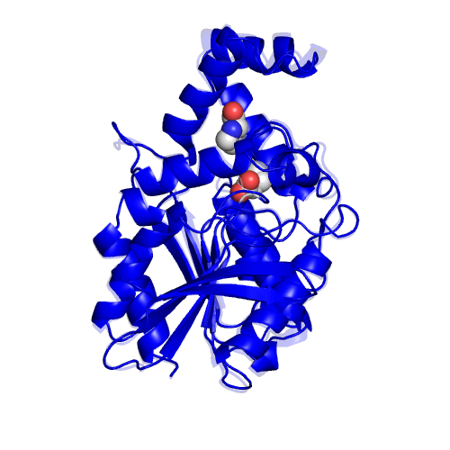
Function |
EC*1 |
CSA distance*2 |
3.4.11.5 |
1.2 |
|
*1 Enzyme commission number. |
Ligand |
PDB*1 |
Full name |
2xPRO |
2x(PROLINE) |
|
*1 Ligand name designated by the PDB identifiers. |
Segments |
Component No. |
Fixed*1 |
Moving*1 |
Motion type |
Ligand binding |
Coupled motion type |
1 |
No significant |
|
*1 The location of the fixed and moving segments indicated by the residue number assigned in the ligand-bound form. The background color of characters indicates the corresponding segment in the structure. The colored segments not described in the Table are: 1) a part of component in which the motion is small (< 1.0 A), or, 2) a part of a protomer of homodimers, for which a corresponding part of the other protomer is shown in the Table. |